

Phlebotomy Order of Draw: Tube Colors & Correct Sequence Guide
August 13, 2026
A blood draw may seem like a simple process, but there is more to it than finding a vein and filling a few tubes. The order in which those tubes are collected can affect the accuracy of the test results.
Why does the order matter? Each tube contains specific additives for different types of laboratory testing. If one tube is collected too early, its additives can carry over into the next tube and interfere with the specimen. That can lead to inaccurate results, a rejected sample, or even a patient needing another blood draw.
That is why the order of draw in phlebotomy is such an important part of safe and accurate venipuncture. It is not just about remembering which color tube comes first. Phlebotomists need to know what each tube contains, what it is used for, and why it belongs at a specific point in the collection process.
What Is the Order of Draw in Phlebotomy?
The order of draw in phlebotomy is the standardized sequence used when collecting multiple blood specimens during a single venipuncture. Each tube may contain a different additive, such as an anticoagulant, clot activator, or glycolytic inhibitor.
Following the correct sequence reduces the risk of additive carryover and helps maintain specimen quality. The same basic order applies whether blood is collected using a tube holder, syringe, or evacuated tube system. Correct sequencing helps with:
- Specimen accuracy: Each sample retains the characteristics required for its intended test.
- Additive control: The sequence reduces unwanted transfer between tubes.
- Reliable laboratory results: Proper collection supports dependable test findings.
- Specimen acceptance: Correct collection can reduce problems that lead to recollection.
- Patient safety: Accurate results help healthcare professionals make appropriate clinical decisions.
Did You Know?
About 70% of laboratory errors occur during the preanalytical phase, which includes steps such as patient identification, specimen collection, tube selection, and sample handling. (Source)
Why Is the Correct Order of Draw Important?
The main reason is additive carryover. If blood from one tube contaminates the next tube with even a small amount of its additive, the specimen may no longer accurately represent the patient’s blood.
For example, anticoagulants used in certain tubes can interfere with testing if they are accidentally carried into another specimen. Using a standardized order helps minimize this risk and supports more reliable laboratory results.
Want to strengthen your practical blood collection skills? Join our Phlebotomy Training Specialists to learn essential phlebotomy techniques and specimen handling.
Standard Phlebotomy Order of Draw and Collection Chart
The standard CLSI sequence for routine venous blood collection is:
| Order | Tube Color | Common Additive | Common Use |
|---|---|---|---|
| 1 | Blood culture | Culture media | Microbiology/blood cultures |
| 2 | Light blue | Sodium citrate | Coagulation testing |
| 3 | Red/Gold | None or clot activator/gel | Serum testing |
| 4 | Green | Heparin | Plasma chemistry testing |
| 5 | Lavender/Purple | EDTA | Hematology testing |
| 6 | Gray | Sodium fluoride/potassium oxalate | Glucose and related testing |
Tube colors can vary by manufacturer and collection system, so the additive and laboratory requirements should always be checked rather than relying on color alone.
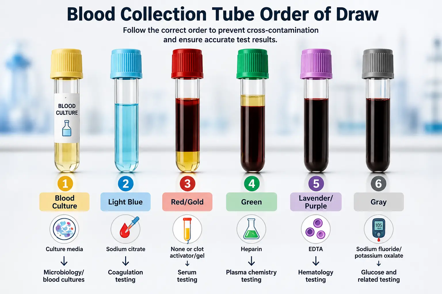
Order of Draw in Phlebotomy: Tube Colors and Sequence
The standard order of draw in phlebotomy moves from blood cultures to citrate, serum, heparin, EDTA, and finally fluoride/oxalate tubes. The sequence is based on the additives inside each tube and the risk that one tube could contaminate the specimen collected after it.
1. Blood Culture Tubes: Collect First
Blood culture specimens come first in the standard venipuncture sequence. They are used to help laboratories detect microorganisms in the bloodstream, making contamination a major concern.
Depending on the collection system, blood cultures may be collected in bottles or specialized culture tubes. Careful skin antisepsis and the facility’s collection procedure are essential because contamination can affect the accuracy of the results.
Key point: Blood culture specimens are collected before tubes containing additives for other tests.
2. Light Blue Tubes: Sodium Citrate
Next come the light blue tubes, which contain sodium citrate and are primarily used for coagulation testing. Common tests include:
- Prothrombin time (PT)
- International normalized ratio (INR)
- Partial thromboplastin time (PTT/aPTT)
- Other coagulation studies
These tubes require the correct blood-to-anticoagulant ratio, so filling the tube to the required level is important. Underfilling can alter the ratio and potentially affect coagulation results. Always follow the manufacturer’s fill requirement and the laboratory’s collection instructions.
3. Red and Gold Tubes: Serum Specimens
After citrate tubes, red and gold tubes are collected for tests requiring serum.
Red tubes may contain no additive or a clot activator, depending on the specific product. Gold tubes commonly contain a clot activator and separator gel. These tubes may be used for a range of chemistry, immunology, and serology tests.
Because tube contents can vary between manufacturers, phlebotomists should confirm the tube requirements for the specific test rather than relying on color alone.
4. Green Tubes: Heparin
The next tubes in the sequence are green heparin tubes. Heparin prevents clotting and allows the laboratory to obtain a plasma specimen for various tests.
Green tubes may be used for selected chemistry, electrolyte, and other plasma-based studies. Different types of heparin tubes have different applications, so checking the tube label and laboratory requirements is important.
5. Lavender and Pink Tubes: EDTA
The following heparin tubes are lavender and pink EDTA tubes. Lavender tubes are widely used for hematology testing, including the complete blood count (CBC). Pink tubes also contain EDTA and are commonly used for blood bank and transfusion-related testing.
EDTA prevents clotting by binding calcium. Because of this, accidental EDTA carryover into another specimen can interfere with tests that require different additives. This is one reason maintaining the correct sequence matters.
6. Gray Tubes: Fluoride/Oxalate
Gray tubes are collected near the end of the standard sequence. They commonly contain sodium fluoride, sometimes combined with an anticoagulant such as potassium oxalate.
These tubes are commonly associated with glucose testing because their additives help limit glycolysis after collection. The exact additive combination can vary, so the tube label and laboratory instructions should always be checked.
Keeping gray tubes toward the end of the sequence helps reduce the risk of their additives carrying over into specimens intended for other tests.
Quick Order of Draw
For routine venipuncture, the sequence can be remembered as:
Blood culture → Light blue → Red/Gold → Green → Lavender/Pink → Gray
The exact tube requirements can vary by manufacturer, test, and laboratory. When in doubt, follow the laboratory’s current collection guidelines rather than relying on tube color alone.
Easy Way to Remember the Order of Draw
A commonly taught sequence is:
Blood Culture → Light Blue → Serum → Green → Lavender → Gray
A simple way to memorize it is to focus on the type of additive rather than memorizing tube colors alone:
Culture → Citrate → Serum → Heparin → EDTA → Fluoride
This approach can make the sequence easier to recall during training and clinical practice.
A systematic review and meta-analysis covering more than 16 million blood sample requests found an overall specimen rejection rate of 1.99%. Clotted specimens (32.23%), hemolysis (22.87%), and insufficient volume (22.81%) were the leading causes of rejection. (Source)

Why Does Each Tube Have a Specific Position?
The sequence in order of draw in phlebotomy relates to the substances inside the tubes. A tube additive can enter the needle or collection pathway and carry into the next specimen. The correct sequence reduces that possibility.
The position of each tube also reflects the type of specimen the laboratory needs. Coagulation testing, serum testing, plasma testing, hematology, and glucose testing can require different additives and handling procedures.
For students, the easiest approach is to learn the sequence alongside the additive and purpose of each tube. Memorizing colors alone can create confusion, especially when specialty tubes enter the collection process.
Building your knowledge beyond tube colors? Learn more about Phlebotomy Technician Certification and take the next step toward developing your professional phlebotomy skills.
Common Order-of-Draw Mistakes to Avoid
Some common mistakes in order-of-draw phlebotomy include:
- Drawing tubes in the wrong sequence
- Relying only on tube color without checking the additive
- Skipping a required tube in a multi-tube collection
- Using a tube that does not match the requested test
- Assuming every manufacturer’s tube uses the same color coding
- Failing to follow facility-specific collection procedures
When a tube or test is not covered by the standard sequence, the laboratory’s collection instructions should take priority. CLSI specifically notes that tubes not included in the standard order should be positioned according to the potential for their additives to affect subsequent specimens.
Does the Order Change With a Butterfly Needle?
Using a butterfly needle does not automatically change the standard order of draw. The sequence remains the same when collecting multiple specimens.
However, special collection considerations may apply depending on the specimen and collection system. For example, when a light-blue coagulation tube is the first tube collected with a butterfly set, a discard tube may be required to ensure the tubing is adequately filled and the correct blood-to-additive ratio is maintained. Follow the laboratory or facility’s specific procedure for these collections.
Tips for Accurate Blood Collection
Correct sequencing is only one part of reliable specimen collection. A phlebotomist also needs to confirm the test requirements, select the correct tube, handle each specimen properly, and complete the collection without avoidable errors.
Keep these practices in mind:
- Verify the test order: Confirm which specimens the laboratory requires before the venipuncture begins.
- Identify the patient correctly: Follow the facility’s approved patient identification procedure before collection.
- Select the correct tubes: Check the tube color, additive, label, and required specimen type.
- Follow the order of draw: Use the approved sequence to reduce the risk of additive carryover.
- Fill tubes correctly: Pay close attention to required fill volumes, especially for coagulation specimens.
- Mix additive tubes properly: Follow the manufacturer’s instructions for gentle inversion and the required number of inversions.
- Avoid vigorous shaking: Excessive force can damage blood cells and compromise specimen quality.
- Label specimens accurately: Complete labeling according to facility requirements before the specimens leave the collection area.
- Check special requirements: Some tests may require specific handling, transport conditions, timing, or tube types.
- Follow facility protocols: When a specialty tube or unusual collection is involved, use the laboratory’s current instructions rather than relying only on memory.
A reliable collection process comes from more than memorizing the sequence. Strong phlebotomy technique combines tube knowledge, careful preparation, accurate identification, and consistent specimen handling.
Ready to take the next step in your phlebotomy career? Learn more about Phlebotomy Technician Certification and the training path that can help prepare you for professional practice.
Keep the Blood Collection Sequence Straight
The correct order of draw in phlebotomy helps protect specimen quality and supports accurate laboratory testing. A simple sequence of blood cultures, light blue, red or gold, green, lavender or pink, and gray tubes gives phlebotomists a reliable foundation for routine venipuncture.
Tube colors alone should not guide every collection. Additives, test requirements, manufacturer instructions, and facility protocols also matter. Strong knowledge of the sequence can help phlebotomy students avoid common collection errors and build confidence during practical phleb tech training.
Frequently Asked Questions
1. What is the correct order of draw in phlebotomy?
The standard order of draw in phlebotomy starts with blood cultures, followed by light blue, red or gold, green, lavender or pink, and gray tubes. Specialty tubes may follow different requirements, so facility instructions should always take priority.
2. Which tube is drawn first in phlebotomy?
Blood culture specimens are generally collected first during routine venipuncture. This position helps reduce the risk of contamination from additives found in other collection tubes.
3. Why is the light blue tube drawn before the red tube?
Light blue tubes contain sodium citrate and support coagulation testing, which requires a controlled blood-to-additive ratio. Collecting them in the correct position helps limit interference from additives in other tubes.
4. What happens if blood tubes are drawn in the wrong order?
An incorrect sequence can allow additives from one tube to enter another specimen and affect test results. The laboratory may identify the problem and request a new specimen when accuracy is compromised.
5. Are phlebotomy tube colors always the same?
Tube colors can vary among manufacturers, laboratories, and specialty collection systems. Phlebotomists should check the tube label, additive, test requirements, and facility protocol instead of relying on color alone.
Latest Posts

August 13, 2024
Is the Role of a CNA the Same as That of a Nurse?

August 13, 2024
Phlebotomists: Vital Contributors to the Healthcare System

August 13, 2024